In some gastrointestinal and pancreatic cancers, the challenge is not reaching the suspicious area, but collecting enough cells to know whether cancer is present. Professor Iyad Khamaysi at Rambam Health Care Campus (Rambam), working alongside scientists from the Technion – Israel Institute of Technology, have developed an innovative solution.
 Professor Iyad Khamaysi. See text for background image information. Photography: Rambam HCC.
Physicians often use brush cytology—a technique in which a small brush is passed over a suspicious area to collect cells for examination under a microscope. However, standard brushes do not always capture or hold enough material for an accurate diagnosis. As a result, tumors may be missed and patients may face delays in treatment.
Professor Khamaysi, director of Rambam's Advanced Endoscopy Unit, director of the Uzia Galil Innovation Center including The Liora & Levy Gerzberg Family Foundation Innovation Laboratory, and Associate Professor at the Technion's Bruce & Ruth Rappaport Faculty of Medicine, worked with Professor Eyal Zussman, head of the Manufacturing Systems Laboratory at the Technion's Faculty of Mechanical Engineering, to take a different approach. Drawing inspiration from the anatomy of a honeybee’s leg, they reimagined the cytology brush, creating a design that could make routine diagnostic sampling more effective.
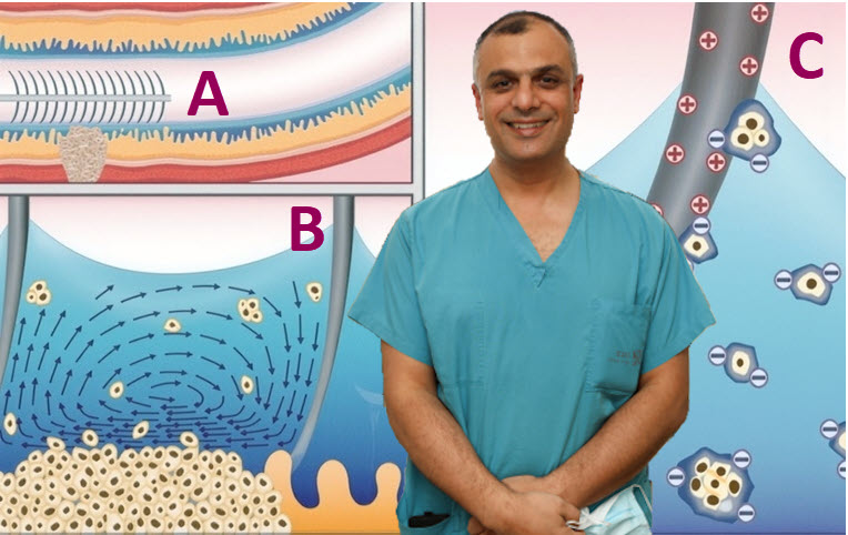
The team decided to enhance the brush itself. They coated its bristles with polyethyleneimine, or PEI, a material that gives the brush a positive surface charge. Since many cancer cells carry a negative charge, the modified brush acts like a magnet, attracting the cells and helping hold them in place. Looking at the featured photo above: A and B) The cytology brush collects cells for diagnosis. C) The positive-charged cytology brush attracts negative-charged cancer cells for analysis.
The research began with a laboratory model designed to better understand how brushing collects cells. Those insights guided the development of the new “smart” brush.
In two studies* published earlier this year, the team demonstrated that, using the same brushing technique, the new device captured twice as many cells in laboratory models and five times more in ex vivo animal models, without changing the way physicians perform the procedure.
Following successful preclinical validation, Professor Khamaysi and Professor Zussman are now preparing for pilot clinical trials at Rambam. Their goal is clear: to make routine screenings more reliable—without increasing cost or risk to patients.
*Read the studes:
